Diagnosis
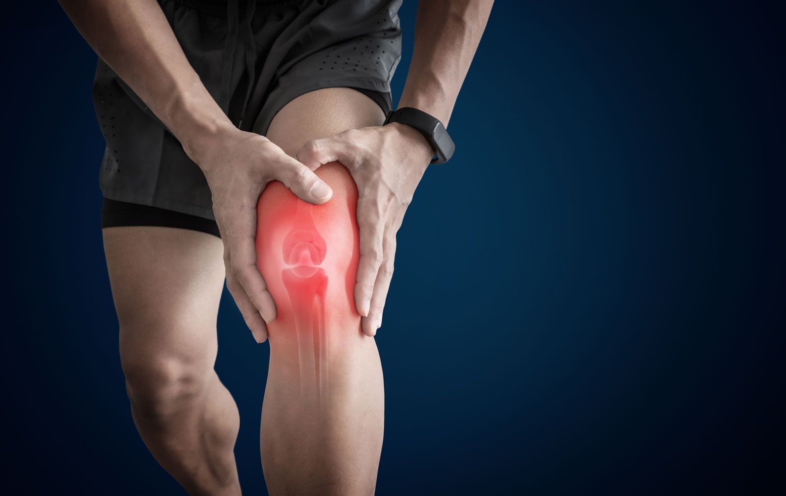
During the physical exam, your doctor will check your joints for swelling, redness, and warmth. He or she will also want to see how well you can move your joints.
Depending on the type of arthritis suspected, your doctor may suggest some of the following tests.
Laboratory tests
The analysis of different types of body fluids can help pinpoint the type of arthritis you may have. Fluids commonly analyzed include blood, urine and joint fluid. To obtain a sample of your joint fluid, your doctor will cleanse and numb the area before inserting a needle in your joint space to withdraw some fluid.
Imaging
These types of tests can detect problems within your joint that may be causing your symptoms. Examples include:
- X-rays. Using low levels of radiation to visualize bone, X-rays can show cartilage loss, bone damage, and bone spurs. X-rays may not reveal early arthritic damage, but they are often used to track the progression of the disease.
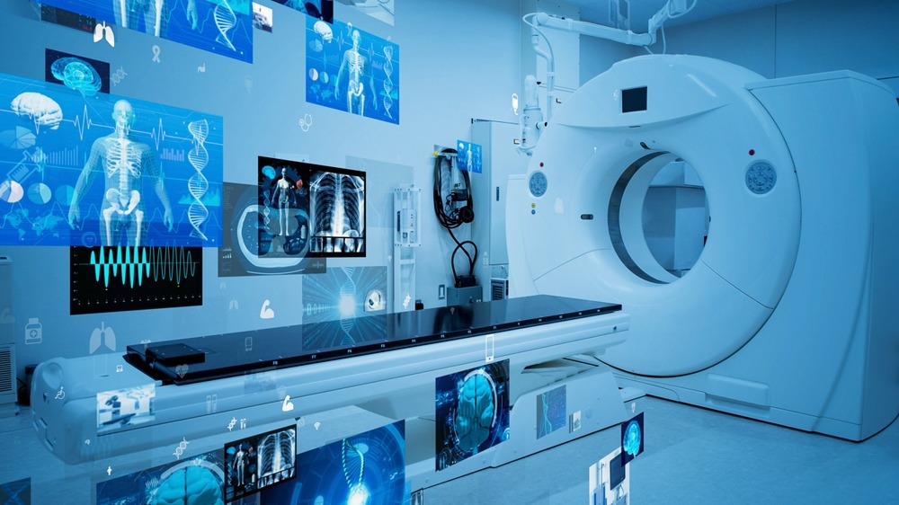
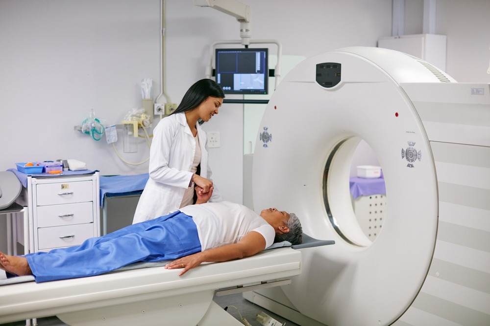
- Computerized tomography (CT). CT scanners take X-rays from many different angles and combine the information to create cross-sectional views of internal structures. CTs can visualize both bone and the surrounding soft tissues.
- Magnetic resonance imaging (MRI). Combining radio waves with a strong magnetic field, MRI can produce more-detailed cross-sectional images of soft tissues such as cartilage, tendons, and ligaments.
- Ultrasound. This technology uses high-frequency sound waves to image soft tissues, cartilage, and fluid-containing structures near the joints (bursae). Ultrasound is also used to guide needle placement for joint aspirations and injections.