enquiry@ecotowndiagnostics.com
Send Your Mail 24/7 Support
Mon - Sat 6:30AM - 10:00PM
Sunday 6:30AM - 3:00PM
Experiencing chest pain can be alarming and often prompts immediate questions about heart health. While chest discomfort doesn’t always signal heart disease, distinguishing between benign causes and potentially serious cardiac issues requires thorough medical evaluation. Among the various diagnostic tools cardiologists use, the Treadmill Tolerance Test, commonly known as TMT or exercise stress test, stands out as a non-invasive yet powerful method for assessing how your heart responds to physical exertion. If you’re experiencing unexplained chest pain and your doctor has recommended cardiac evaluation, understanding what a treadmill test in Bangalore involves and how it detects heart blockages can help you approach the examination with confidence and clarity.
The TMT test represents a cornerstone of cardiac diagnostics, providing cardiologists with crucial information about coronary artery function, exercise capacity, and heart rhythm abnormalities. This comprehensive examination goes beyond resting electrocardiograms by revealing problems that only become apparent when the heart works harder during physical activity.
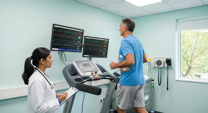
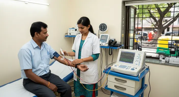
The Treadmill Tolerance Test monitors your heart’s electrical activity, blood pressure, and symptoms while you walk on a treadmill test in Bangalore at progressively increasing speeds and inclines. This controlled stress environment reveals how well your coronary arteries supply blood to your heart muscle during increased demand, making it an invaluable tool for detecting blockages and assessing overall cardiovascular fitness.
During rest, partially blocked coronary arteries may supply adequate blood flow to meet your heart’s minimal oxygen requirements. However, when you exercise, your heart muscle demands significantly more oxygen-rich blood. If coronary blockages restrict blood flow, the heart muscle downstream from the blockage doesn’t receive sufficient oxygen—a condition called ischemia. This oxygen deficit triggers changes in the heart’s electrical activity visible on the electrocardiogram and often causes chest discomfort or other symptoms.
By progressively increasing exercise intensity, the TMT test gradually elevates your heart’s oxygen demand until reaching your target heart rate or until symptoms develop. This systematic approach helps identify coronary artery disease that remains silent during ordinary daily activities but emerges during physical exertion.
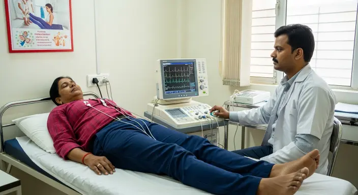
Throughout the examination, medical professionals continuously monitor multiple parameters that collectively provide comprehensive information about cardiac function. The electrocardiogram records your heart’s electrical signals through electrodes placed on your chest, revealing changes in the ST segment—the portion of the heartbeat cycle most sensitive to ischemia. Specific ST segment changes indicate insufficient blood flow to heart muscle regions.
Blood pressure measurements taken at regular intervals reveal how your cardiovascular system responds to increasing workload. Normally, systolic blood pressure rises with exercise intensity while diastolic pressure remains stable or decreases slightly. Abnormal responses—such as blood pressure failing to rise appropriately or dropping during exercise—can indicate significant cardiac problems.
Heart rate monitoring ensures you reach the target heart rate appropriate for your age, calculated as approximately 85 percent of your maximum predicted heart rate. Achieving this target maximizes the test’s diagnostic sensitivity by ensuring adequate cardiac stress.
Cardiologists don’t recommend TMT testing for everyone experiencing chest discomfort. Understanding when this examination provides valuable diagnostic information helps explain why your doctor might suggest it or choose alternative tests.
Chest pain arising from coronary artery disease typically presents with specific characteristics that distinguish it from non-cardiac causes. Angina—chest pain caused by inadequate blood flow to heart muscle—typically feels like pressure, squeezing, heaviness, or tightness in the chest center or left side. The discomfort often radiates to the left arm, neck, jaw, or back.
Cardiac chest pain typically occurs during physical exertion or emotional stress when the heart requires increased blood flow, and it improves with rest as oxygen demand decreases. Episodes usually last several minutes rather than fleeting seconds or continuous hours. If your chest pain follows these patterns, your doctor will likely recommend cardiac evaluation including possible TMT testing.
Even if your chest pain doesn’t perfectly match classic angina patterns, having multiple cardiovascular risk factors increases the likelihood that your symptoms reflect underlying heart disease. Risk factors include diabetes, high blood pressure, elevated cholesterol, smoking history, family history of early heart disease, obesity, and sedentary lifestyle.
The combination of chest pain and significant risk factors creates moderate pretest probability for coronary artery disease—the ideal scenario for TMT testing. When disease likelihood is moderate, exercise stress testing provides valuable diagnostic information that helps guide further management.
Sometimes physicians recommend TMT testing to reassure patients and exclude cardiac causes when chest pain characteristics suggest low likelihood of coronary disease but uncertainty remains. A normal stress test result in someone with atypical chest pain and few risk factors provides reassurance that serious heart disease is unlikely, allowing focus on alternative explanations for symptoms.
Proper preparation ensures optimal test quality and your safety during the examination. Understanding what to do before arriving at the testing facility helps the procedure go smoothly.
Your cardiologist provides specific instructions about medications before testing. Beta-blockers and certain other cardiac medications may need to be temporarily discontinued because they can affect heart rate response and potentially mask significant findings. Never stop medications without explicit physician approval, as some cardiac drugs require gradual tapering to avoid rebound effects.
Bring a complete list of all medications including dosages to your appointment. If you take insulin or diabetes medications, discuss timing with your physician since fasting before the test affects blood sugar levels.
Wear comfortable athletic shoes with good support and loose-fitting clothing that allows free movement. Women should wear a sports bra or comfortable top since electrodes will be placed on the chest. Avoid applying lotions, oils, or powders to your chest area, as these interfere with electrode adhesion.
Plan to be at the testing facility for approximately two hours, though the actual treadmill test in Bangalore walking typically lasts only 8 to 12 minutes. The additional time accommodates preparation, monitoring, and post-exercise observation.
Most facilities request that you avoid eating for three to four hours before testing to prevent nausea during exercise. Light meals several hours beforehand are acceptable. Continue drinking water to maintain adequate hydration.
Avoid vigorous exercise for 24 hours before testing to ensure you can perform maximally during the actual examination. Fatigue from prior workouts might prevent reaching target heart rate, reducing diagnostic accuracy.
Understanding what happens during the examination helps reduce anxiety and ensures you can cooperate effectively with the medical team’s instructions.
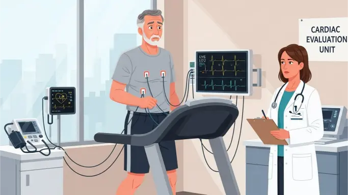
Upon arrival, you complete questionnaires about medical history and current symptoms. The technician explains the procedure and obtains informed consent. Electrode placement requires shaving small patches of chest hair if necessary to ensure good electrical contact. Typically, ten electrodes are positioned at specific locations on your chest, arms, and legs.
Baseline measurements include a resting electrocardiogram and blood pressure while you stand quietly. These resting values provide comparison points for changes occurring during exercise.
You begin walking on the treadmill at a slow, comfortable pace on a flat surface. Every three minutes, the protocol increases both speed and incline according to standardized stages called the Bruce protocol. Most people complete several stages before reaching their target heart rate or developing limiting symptoms.
The medical team continuously monitors your electrocardiogram, observes for symptoms, and measures blood pressure every few minutes. You’ll be asked repeatedly about chest discomfort, shortness of breath, leg fatigue, or other symptoms. Being honest about symptoms is crucial for your safety and test accuracy.
The test continues until you reach target heart rate, develop significant symptoms, show concerning electrocardiogram changes, experience blood pressure abnormalities, or reach exhaustion. You can request to stop at any time, though maximal effort provides the most diagnostic information.
Immediately after stopping exercise, you continue walking slowly for several minutes during the cool-down phase. This gradual reduction in activity helps prevent blood pooling in your legs and reduces the risk of post-exercise lightheadedness. The medical team continues monitoring your electrocardiogram and blood pressure closely during recovery since some cardiac changes appear only after exercise ends.
You remain under observation until your heart rate and blood pressure approach baseline values and any symptoms resolve. Most people feel completely normal within 10 to 15 minutes after finishing the treadmill test in Bangalore portion.
Understanding how exercise stress testing reveals coronary artery blockages helps you appreciate what the examination accomplishes and why certain findings raise concern.
The hallmark of significant coronary blockage during TMT testing is ST segment depression on the electrocardiogram. When heart muscle doesn’t receive adequate oxygen during exercise, the affected cells generate altered electrical signals visible as horizontal or downsloping ST segment depression measuring at least one millimeter.
The location of ST segment changes sometimes indicates which coronary artery has blockages, though this correlation isn’t perfectly reliable. The magnitude and duration of changes provide information about ischemia severity.
Chest pain, pressure, or discomfort developing during exercise and resolving with rest strongly suggests angina caused by coronary blockages. The exercise intensity at which symptoms appear provides information about disease severity—symptoms occurring at low workloads suggest more significant blockages than symptoms appearing only with maximal exertion.
Other concerning symptoms include unusual shortness of breath disproportionate to exercise intensity, lightheadedness, or extreme fatigue. These manifestations sometimes indicate significant cardiac dysfunction even without typical chest pain.
Failure of blood pressure to rise appropriately during exercise can indicate severe coronary disease or impaired heart muscle function. Similarly, blood pressure dropping during exercise represents a concerning finding suggesting significant cardiac compromise.
Inappropriate heart rate responses, though less specific than other findings, sometimes indicate chronotropic incompetence—the heart’s inability to increase rate appropriately with exercise demand.
Understanding what constitutes normal versus abnormal results helps you comprehend your test findings and their implications for your cardiac health.
A negative or normal TMT test means you achieved adequate exercise intensity without developing electrocardiogram changes, symptoms, or blood pressure abnormalities suggesting cardiac ischemia. This finding provides reassurance that significant coronary blockages are unlikely, especially if you reached good exercise capacity.
However, stress testing isn’t perfectly sensitive—some patients with coronary disease have normal stress tests, particularly if blockages aren’t severe enough to cause ischemia at achieved exercise levels. Your doctor interprets results in the context of your overall clinical picture.
A positive TMT test indicates findings suggestive of coronary artery disease. The specific abnormalities detected, combined with when during exercise they appeared and their severity, help risk-stratify patients. Strongly positive tests showing marked electrocardiogram changes, significant symptoms, or abnormalities at low exercise workloads suggest high-risk disease requiring prompt further evaluation.
Positive stress tests typically lead to additional testing, most commonly coronary angiography, to definitively identify blockage locations and severity. This invasive test threads a catheter into coronary arteries and injects contrast dye, creating detailed pictures showing exactly where and how severely arteries are blocked.
Sometimes TMT testing yields equivocal results due to submaximal heart rate achievement, non-diagnostic electrocardiogram changes, or inability to complete adequate exercise. These inconclusive tests don’t provide definitive answers about coronary disease presence or absence.
When results are equivocal, your doctor might recommend alternative stress testing methods like pharmacologic stress testing with imaging, stress echocardiography, or coronary CT angiography to obtain clearer diagnostic information.
Despite newer cardiac imaging technologies, TMT testing remains valuable due to several distinct advantages that make it appropriate as a first-line diagnostic tool for many patients.
The TMT test requires no injections, radiation exposure, or invasive procedures. The examination uses readily available, relatively inexpensive equipment found in most cardiac facilities. This widespread availability makes it accessible to patients throughout urban and suburban areas.
Unlike anatomic imaging that simply shows whether blockages exist, exercise stress testing provides functional information about whether blockages cause actual ischemia during physical activity. Some anatomically significant blockages don’t cause functional limitation, while some moderate blockages do produce ischemia. The TMT test answers the clinically relevant question: does your heart receive adequate blood flow during exertion?
Beyond detecting ischemia, the test provides valuable information about your overall exercise capacity and functional status. The maximum stage achieved and exercise duration correlate with cardiovascular fitness and have prognostic significance independent of ischemia detection. Poor exercise capacity predicts increased cardiovascular risk even when electrocardiogram changes don’t occur.
TMT test results help stratify cardiovascular risk, guiding decisions about medical management intensity and need for invasive procedures. Low-risk test results support conservative medical management, while high-risk findings prompt aggressive intervention.
While TMT testing provides valuable information for many patients, certain situations limit its usefulness or make alternative tests more appropriate.
Patients with certain baseline electrocardiogram abnormalities—including left bundle branch block, paced rhythms, or ST segment changes at rest—cannot undergo interpretable TMT testing because these conditions obscure ischemic changes. For these individuals, stress testing with imaging (nuclear or echocardiographic) provides more accurate information.
Orthopedic problems, neurological conditions, severe deconditioning, or peripheral vascular disease may prevent adequate treadmill exercise. Patients unable to walk on a treadmill require alternative testing methods, typically pharmacologic stress tests using medications to increase heart rate or dilate coronary arteries without physical exercise.
Exercise stress testing has somewhat lower accuracy in women compared to men, with higher rates of false-positive results. Some cardiologists prefer stress testing with imaging for female patients, particularly those with multiple risk factors or concerning symptoms.
Understanding what happens after TMT testing helps you prepare for potential follow-up procedures or treatment recommendations.
If your stress test is normal and your symptoms are atypical for cardiac disease, your doctor typically reassures you that significant coronary disease is unlikely. Attention shifts to identifying alternative explanations for chest discomfort, which might include musculoskeletal problems, gastrointestinal issues, or anxiety-related symptoms.
Even with normal results, your physician addresses cardiovascular risk factors through lifestyle modifications and medications when appropriate to prevent future heart disease development.
Positive stress test results usually prompt coronary angiography to definitively visualize coronary arteries and identify blockage locations and severity. Angiography findings determine whether you need medical management alone, percutaneous coronary intervention with stent placement, or coronary artery bypass surgery.
Some patients with mildly positive stress tests and low-risk features might undergo non-invasive coronary CT angiography instead of invasive catheterization, depending on clinical circumstances and physician preference.
While standard TMT testing provides valuable information, enhanced protocols combining exercise with imaging techniques offer additional diagnostic capabilities for specific clinical situations.
Stress echocardiography combines treadmill test in Bangalore exercise with ultrasound imaging of the heart performed immediately after peak exercise. This technique visualizes heart wall motion abnormalities that develop when coronary blockages cause ischemia. Stress echocardiography offers higher diagnostic accuracy than electrocardiogram monitoring alone.
Nuclear stress tests inject small amounts of radioactive tracer into your bloodstream during peak exercise. Special cameras create images showing tracer distribution in heart muscle, revealing areas receiving inadequate blood flow. This technique provides excellent diagnostic accuracy and helps localize ischemia to specific coronary artery territories.
When facing chest pain evaluation, understanding the role of exercise stress testing empowers you to participate actively in your cardiac care. The TMT test provides valuable functional information about how your heart responds to physical demands, revealing blockages that might not cause symptoms at rest but become problematic during exertion. This non-invasive examination offers an excellent balance of diagnostic accuracy, safety, and accessibility for many patients requiring cardiac evaluation.
While no test is perfect, the TMT examination has proven its value over decades of clinical use. Modern cardiac care often combines stress testing results with other clinical information, risk factor assessment, and sometimes additional imaging to create comprehensive pictures of cardiovascular health. Whether your test reveals reassuring normal function or identifies concerning abnormalities requiring further evaluation, the information obtained guides appropriate management strategies tailored to your specific situation.
Exercise stress testing remains a cornerstone of chest pain evaluation despite impressive advances in cardiac imaging technology. The TMT test’s ability to assess functional cardiac capacity, detect exercise-induced ischemia, and stratify cardiovascular risk makes it an invaluable diagnostic tool for appropriate patient populations. When you undergo a treadmill test in Bangalore for chest pain evaluation, you’re receiving a time-tested cardiac assessment that provides crucial information about coronary artery function and overall heart health.
Understanding what the examination measures, how it detects blockages, and what results mean for your cardiac care helps you approach the test with confidence. Whether results reveal normal cardiac function providing reassurance or identify concerns requiring additional evaluation, the TMT test represents an essential step in comprehensive cardiovascular assessment, helping ensure you receive appropriate treatment tailored to your heart’s specific needs and ultimately supporting the best possible cardiovascular outcomes.