The human heart beats approximately 100,000 times each day, pumping blood through an intricate network of vessels that extends over 60,000 miles throughout the body. When something goes wrong with this remarkable system, early detection becomes crucial for successful treatment and management. Modern medical imaging has revolutionized cardiovascular diagnostics, and among these innovations, Doppler ultrasound stands out as a powerful, non-invasive tool. For those seeking reliable diagnostic services, finding a quality Doppler scan in Bangalore has become increasingly accessible, offering patients advanced technology and expert interpretation.
Understanding Doppler Ultrasound Technology
Doppler ultrasound represents a significant advancement over traditional ultrasound imaging. While conventional ultrasound creates images of structures within the body, Doppler technology adds an extra dimension by measuring the movement of blood through vessels and heart chambers. This technique employs the Doppler effect, the same principle that explains why an ambulance siren’s pitch changes as it passes by.
When sound waves bounce off moving blood cells, their frequency shifts. The ultrasound machine detects these changes and converts them into detailed information about blood flow direction, speed, and patterns. This real-time visualization allows physicians to spot abnormalities that might indicate serious cardiovascular conditions long before symptoms become severe.
Early Detection of Heart Valve Problems Through Doppler Scan in Bangalore
Heart valves function as one-way gates, ensuring blood flows in the correct direction through the heart’s chambers. When these valves malfunction, either narrowing (stenosis) or leaking (regurgitation), the heart must work harder to maintain adequate circulation. Doppler ultrasound excels at identifying these valve disorders in their earliest stages.
The technology can detect even subtle changes in blood flow patterns across heart valves. Medical professionals can measure the pressure gradients across valves, assess the severity of leaks, and determine whether surgical intervention might be necessary. Early identification through advanced diagnostic centers offering Doppler scan in Bangalore enables patients to begin treatment before permanent heart damage occurs.
Recognizing Valve Stenosis
Valve stenosis occurs when heart valves become stiff and narrow, restricting blood flow. Doppler imaging reveals increased blood flow velocity through narrowed valves, helping cardiologists grade the severity of stenosis. Conditions like aortic stenosis, which can progress silently for years, become visible through Doppler examination long before chest pain or fainting episodes develop.
Identifying Valve Regurgitation
When heart valves fail to close completely, blood leaks backward through the valve. Doppler technology creates color-coded images showing this abnormal reverse flow, called regurgitation. Physicians can quantify the volume of leaking blood and monitor progression over time, adjusting treatment strategies as needed.
Detecting Coronary Artery Disease and Blood Flow Obstructions
Coronary artery disease remains one of the leading causes of death worldwide, yet early stages often produce no symptoms. Doppler ultrasound provides a window into coronary blood flow, revealing areas where arterial narrowing reduces oxygen supply to heart muscle. While coronary angiography remains the gold standard for visualizing coronary arteries, Doppler studies offer valuable screening information without invasive procedures.
The technology proves particularly valuable in assessing peripheral arterial disease, where blood flow to limbs becomes compromised. Patients experiencing leg pain, numbness, or wounds that heal slowly may benefit from Doppler examination. The test identifies blockages or narrowing in arteries supplying the legs, arms, or other regions, guiding treatment decisions that can prevent tissue loss or amputation.
Evaluating Congenital Heart Defects
Some individuals are born with structural heart abnormalities that may not become apparent until later in life. Doppler ultrasound plays a crucial role in diagnosing these congenital defects, from simple holes between heart chambers to complex malformations affecting multiple structures.
The technology reveals abnormal blood flow patterns characteristic of conditions like atrial septal defects, ventricular septal defects, and patent ductus arteriosus. Early detection through comprehensive screening allows for timely intervention, often preventing complications that could affect quality of life or longevity.
Assessing Heart Muscle Function with Doppler Scan in Bangalore
Beyond valves and blood vessels, Doppler technology evaluates how effectively the heart muscle contracts and relaxes. This assessment proves vital for diagnosing cardiomyopathies, conditions where heart muscle becomes enlarged, thick, or rigid. These disorders can lead to heart failure if left undetected and untreated.
Doppler measurements reveal whether the heart fills properly during relaxation (diastolic function) and whether it pumps efficiently during contraction (systolic function). Subtle abnormalities in these measurements often precede noticeable symptoms, offering a critical window for intervention. Modern facilities providing Doppler scan in Bangalore utilize advanced equipment capable of detecting even minor functional impairments.
Monitoring Deep Vein Thrombosis and Pulmonary Embolism Risk
Blood clots within deep veins, particularly in the legs, pose serious health risks. When these clots break free and travel to the lungs, they can cause life-threatening pulmonary embolism. Doppler ultrasound serves as the primary diagnostic tool for detecting deep vein thrombosis, visualizing blood flow obstruction caused by clots.
The examination proves especially important for individuals with risk factors such as prolonged immobility, recent surgery, cancer, or pregnancy. Quick diagnosis through accessible services like Doppler scan in Bangalore enables prompt anticoagulation therapy, preventing clot progression and reducing embolism risk.
Understanding Carotid Artery Disease
The carotid arteries supply blood to the brain, and blockages in these vessels significantly increase stroke risk. Doppler ultrasound of the carotid arteries identifies atherosclerotic plaque buildup and measures the degree of arterial narrowing. This non-invasive screening proves invaluable for stroke prevention, as many patients with significant carotid disease experience no warning symptoms.
The technology differentiates between stable and unstable plaque, helping physicians assess stroke risk more accurately. Regular monitoring through Doppler studies allows doctors to track disease progression and determine optimal timing for interventions ranging from medication management to surgical procedures.
The Advantages of Non-Invasive Cardiovascular Assessment
Traditional cardiovascular diagnostics often required invasive procedures involving catheter insertion into blood vessels. While these techniques remain necessary for certain situations, Doppler ultrasound offers comparable information without needles, incisions, or radiation exposure. The painless procedure typically takes less than an hour and carries virtually no risk of complications.
Patients can undergo repeated examinations as needed for disease monitoring without concern about cumulative radiation exposure or procedural risks. This safety profile makes Doppler studies ideal for screening high-risk populations, following treatment response, and conducting long-term surveillance of chronic conditions. The widespread availability of quality Doppler scan in Bangalore ensures that residents have access to this essential diagnostic service.
Applications Beyond Cardiac Assessment
While cardiovascular applications dominate Doppler ultrasound usage, the technology extends to other circulatory disorders. Physicians use Doppler studies to evaluate blood flow in transplanted organs, assess fetal circulation during pregnancy, diagnose varicoceles affecting male fertility, and investigate causes of erectile dysfunction related to blood flow problems.
The versatility of this imaging modality continues to expand as technical capabilities improve. Three-dimensional Doppler imaging and contrast-enhanced studies push the boundaries of what clinicians can visualize and measure, opening new possibilities for early disease detection and treatment monitoring.
Preparing for Your Doppler Ultrasound Examination
Most Doppler ultrasound examinations require minimal preparation. Patients typically wear comfortable clothing and may need to remove jewelry or accessories that could interfere with imaging. For abdominal vascular studies, fasting for several hours before the examination might be recommended to reduce intestinal gas that can obscure vessel visualization.
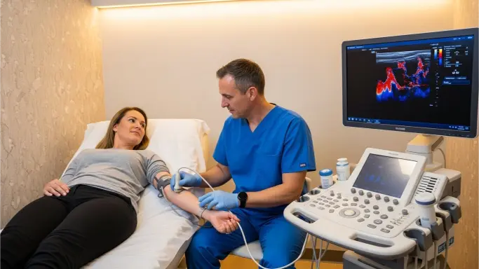
During the procedure, a technician applies gel to the skin and moves a handheld transducer over the area being examined. The device sends and receives sound waves, creating real-time images displayed on a monitor. Patients may hear whooshing sounds representing blood flow, and the technician might ask them to breathe deeply, hold their breath, or change positions to optimize imaging.
The Future of Doppler Technology in Cardiovascular Medicine
Ongoing technological advancement continues enhancing Doppler ultrasound capabilities. Artificial intelligence algorithms now assist in image interpretation, potentially improving diagnostic accuracy and consistency. Portable Doppler devices bring sophisticated cardiovascular assessment to emergency rooms, ambulances, and remote locations where traditional imaging equipment isn’t available.
Integration with other imaging modalities creates comprehensive cardiovascular evaluation platforms. Research explores novel applications for Doppler technology, from assessing myocardial microcirculation to detecting vulnerable atherosclerotic plaques before they rupture and cause heart attacks or strokes.
Choosing Quality Diagnostic Services
When seeking cardiovascular assessment, the quality of equipment and expertise of interpreting physicians significantly impact diagnostic accuracy. Modern facilities offering Doppler scan in Bangalore typically feature state-of-the-art ultrasound systems capable of advanced imaging modes and precise measurements. Board-certified cardiologists or radiologists with specialized training in vascular ultrasound should interpret study results to ensure accurate diagnosis.
Patients should feel comfortable asking about technician credentials, physician qualifications, and equipment specifications when selecting a diagnostic center. Facilities committed to quality maintain accreditation from recognized professional organizations and regularly update their technology to reflect current best practices.
Conclusion
The ability to detect heart and circulatory disorders before they produce symptoms or cause irreversible damage represents one of modern medicine’s greatest achievements. Doppler ultrasound technology stands at the forefront of this early detection capability, offering safe, accurate, and accessible cardiovascular assessment. From valve disorders and coronary disease to blood clots and congenital defects, this versatile imaging modality reveals problems that might otherwise remain hidden until they reach critical stages.
For individuals in urban areas seeking reliable cardiovascular screening, services providing Doppler scan in Bangalore offer comprehensive diagnostic capabilities backed by advanced technology and skilled medical professionals. Regular cardiovascular assessment through Doppler ultrasound, particularly for those with risk factors, can truly save lives by identifying treatable conditions before they progress to emergencies. As technology continues advancing and awareness grows, Doppler ultrasound will undoubtedly play an increasingly vital role in preventive cardiovascular care and early disease management.