Pregnancy brings countless moments of anticipation and wonder, but it also requires careful monitoring to ensure both mother and baby remain healthy throughout the journey. Among the various prenatal examinations, growth scans play a crucial role in tracking fetal development and identifying potential concerns early. If you’re expecting and have been recommended for additional monitoring, understanding what growth scans entail and when they become necessary can help you feel more confident about your prenatal care. When seeking an ultrasound scan in Bangalore, knowing the purpose and timing of these specialized examinations ensures you receive appropriate care tailored to your pregnancy needs.
Growth scans, also called fetal biometry scans, represent specialized ultrasound examinations designed to assess your baby’s physical development at various stages of pregnancy. Unlike routine prenatal ultrasounds that check overall wellbeing, these targeted scans focus specifically on measuring fetal size and comparing those measurements against expected growth patterns for gestational age.
Understanding What Growth Scans Actually Measure
Growth scans utilize sophisticated ultrasound scan in Bangalore technology to obtain precise measurements of specific fetal body parts. These measurements collectively provide comprehensive information about your baby’s development and help healthcare providers identify any deviations from normal growth trajectories.
Key Measurements Taken During Growth Scans
The primary measurement obtained during every growth scan is the biparietal diameter, which measures the width of your baby’s head from one side to the other. This measurement provides crucial information about brain development and helps establish gestational age when combined with other parameters.
Head circumference represents another vital measurement, calculated by measuring around the entire perimeter of your baby’s skull. This measurement complements the biparietal diameter and provides additional insight into cranial and brain growth patterns.
Abdominal circumference measures the distance around your baby’s belly at the level of the liver and stomach. This measurement proves particularly valuable because it reflects nutritional status and overall body growth. Babies not receiving adequate nutrition often show reduced abdominal circumference before other measurements are affected.
Femur length measures the longest bone in your baby’s thigh, providing information about skeletal development. This measurement helps assess overall body length and proportionality when compared with other body measurements.
Calculating Estimated Fetal Weight
Sonographers use sophisticated formulas that incorporate multiple measurements to calculate estimated fetal weight. While no formula provides perfectly accurate predictions, modern calculations typically estimate weight within 10 to 15 percent of actual birth weight. This estimation helps physicians determine whether your baby’s size is appropriate for gestational age.
Understanding that estimated fetal weight represents an approximation rather than an exact figure helps manage expectations. Factors like maternal body habitus, fetal position, and amniotic fluid volume can influence measurement accuracy.
When Healthcare Providers Recommend Growth Scans
Not every pregnancy requires additional growth scanning beyond the standard anatomy scan performed around 20 weeks gestation. Healthcare providers recommend these specialized examinations when specific risk factors or clinical situations suggest closer monitoring would benefit maternal and fetal health.
High-Risk Pregnancy Conditions
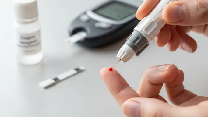
Women with pre-existing medical conditions like diabetes or hypertension typically receive more frequent growth monitoring. These conditions can affect placental function and consequently impact fetal growth patterns. Regular scanning allows early detection of growth restriction or excessive growth, enabling timely intervention.
Pregnancy-induced hypertension or preeclampsia raises concerns about placental insufficiency, which can compromise nutrient and oxygen delivery to the developing baby. Growth scans help monitor whether these conditions are affecting fetal development.
Previous Pregnancy Complications
If you experienced intrauterine growth restriction, preterm delivery, or stillbirth in previous pregnancies, your healthcare provider will likely recommend serial growth scans. These past experiences indicate elevated risk for similar complications, making proactive monitoring essential.
Women who previously delivered babies significantly larger or smaller than expected often receive additional scanning in subsequent pregnancies to watch for recurring patterns.
Understanding Growth Patterns and Percentiles Through Ultrasound Scan in Bangalore
When you receive results from growth scans, measurements are typically expressed as percentiles that compare your baby’s size to populations of babies at the same gestational age. Understanding what these percentiles mean helps interpret whether your baby’s growth pattern raises concerns.
Normal Growth Ranges
Babies measuring between the 10th and 90th percentiles for their gestational age are generally considered appropriately grown. This range encompasses the vast majority of healthy babies and indicates development is proceeding normally.
The 50th percentile represents the average or median size for a specific gestational age. Babies at this percentile are exactly average-sized compared to the reference population.
Small for Gestational Age Concerns
When measurements consistently fall below the 10th percentile, babies are classified as small for gestational age. This classification doesn’t automatically indicate problems, as some babies are constitutionally small due to genetic factors. However, it triggers closer monitoring to distinguish between healthy small babies and those experiencing true growth restriction.
Intrauterine growth restriction represents a more serious condition where the baby fails to achieve expected growth potential due to placental insufficiency, maternal health issues, or fetal problems. Serial scans tracking growth velocity over time help distinguish growth restriction from constitutional smallness.
Large for Gestational Age Considerations
Babies measuring above the 90th percentile are classified as large for gestational age. While many large babies are perfectly healthy, this classification can indicate gestational diabetes or other maternal conditions affecting fetal growth. Large babies face increased risks for birth complications, making accurate size assessment important for delivery planning.
Additional Assessments During Growth Scans
Comprehensive growth examinations extend beyond simple measurements to evaluate other important aspects of fetal wellbeing and the intrauterine environment.
Amniotic Fluid Volume Assessment
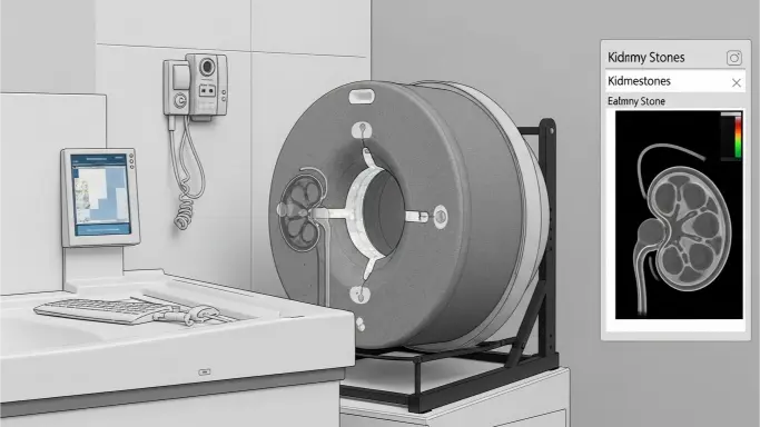
Sonographers measure amniotic fluid volume during growth scans because fluid levels provide indirect information about fetal kidney function and placental health. Too little fluid, called oligohydramnios, may indicate placental insufficiency or fetal kidney problems. Excessive fluid, or polyhydramnios, can suggest gestational diabetes or fetal swallowing difficulties.
The amniotic fluid index divides the uterus into quadrants and measures the deepest pocket of fluid in each section. Alternatively, the single deepest pocket method measures only the largest fluid collection. Both approaches help determine whether fluid volume falls within normal ranges.
Placental Location and Appearance
Growth scans document placental position to identify concerning locations like placenta previa, where the placenta covers the cervical opening. Sonographers also assess placental texture and appearance, looking for signs of premature aging or structural abnormalities that might affect function.
Umbilical Cord and Blood Flow
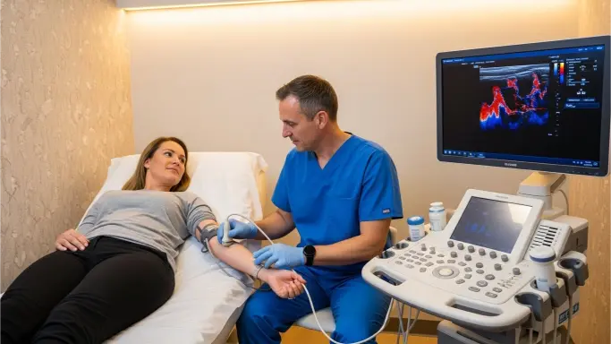
Evaluation of the umbilical cord includes counting vessels—normally two arteries and one vein—and checking for abnormalities in cord insertion or structure. Some centers include Doppler studies during growth scans to assess blood flow through the umbilical cord and fetal vessels, providing information about placental function and fetal adaptation to the intrauterine environment.
Frequency and Timing of Growth Monitoring
The schedule for growth scans varies based on individual risk factors and clinical indications. Understanding typical timing helps you plan for these important appointments.
Standard Growth Scan Timing
When growth monitoring becomes necessary, scans typically begin around 28 to 32 weeks gestation and continue every 2 to 4 weeks until delivery. This timing allows adequate intervals for meaningful growth to occur between examinations while providing regular updates on fetal development.
More frequent scanning may be recommended if significant concerns arise or if measurements show worrisome trends. Conversely, if initial growth scans show reassuring patterns in low-risk situations, providers might space examinations further apart.
Third Trimester Focus
Most growth concerns become apparent during the third trimester when fetal growth accelerates rapidly. This period represents the time when placental insufficiency or maternal conditions most commonly affect fetal development, making it the primary window for growth monitoring.
Some conditions warrant earlier initiation of growth scanning, particularly if ultrasound scan in Bangalore findings or maternal symptoms suggest problems developing before the third trimester.
What Happens If Growth Issues Are Detected
Discovering growth abnormalities doesn’t automatically mean serious problems exist, but it does trigger additional evaluation and monitoring to ensure optimal outcomes.
Enhanced Surveillance Protocols
When growth restriction is identified, healthcare providers typically implement enhanced surveillance protocols including more frequent growth scans, non-stress tests to assess fetal heart rate patterns, and biophysical profiles evaluating fetal movement, breathing, and amniotic fluid.
This comprehensive monitoring helps determine whether the baby is tolerating the intrauterine environment well or showing signs of compromise that might necessitate early delivery.
Lifestyle and Medical Interventions
Some growth concerns respond to specific interventions. Women with inadequate nutrition may benefit from dietary counseling. Those with uncontrolled maternal conditions require optimization of medical management. Bed rest or activity modification might be recommended in certain situations, though evidence supporting these interventions varies.
Delivery Planning Considerations
Growth scan findings significantly influence delivery planning. Babies identified as large for gestational age might prompt discussions about delivery timing or cesarean delivery if vaginal birth appears risky. Growth-restricted babies may require early delivery if the intrauterine environment becomes less favorable than the neonatal intensive care unit.
The decision to deliver balances risks of prematurity against risks of continued pregnancy, with growth scan trends providing crucial information for this complex calculation.
Accuracy and Limitations of Growth Scanning
While growth scans provide valuable information, understanding their limitations helps maintain realistic expectations about what these examinations can and cannot tell you.
Measurement Variability
Even with experienced sonographers and high-quality equipment, measurements contain inherent variability. Different operators may obtain slightly different measurements, and fetal position significantly affects measurement accuracy. This variability explains why providers focus on growth trends over time rather than single measurements when making clinical decisions.
Weight Estimation Imperfections
Estimated fetal weight calculations, while useful, lack perfect precision. Errors of 10 to 15 percent are common, meaning a baby estimated to weigh 2500 grams might actually weigh anywhere from 2125 to 2875 grams at birth. This imprecision is important to remember when discussing size-related concerns or delivery planning.
Constitutional Variation
Not every baby measuring small or large has an underlying problem. Genetic factors, parental size, ethnicity, and other variables create normal variations in fetal size. Distinguishing healthy constitutional variation from pathological growth abnormalities requires clinical judgment incorporating multiple factors beyond scan measurements alone.
Technology Advances Improving Growth Assessment
Modern ultrasound technology continues evolving, improving the accuracy and comprehensiveness of fetal growth evaluation.
Three-Dimensional Imaging
Three-dimensional ultrasound scan in Bangalore creates volumetric images of the fetus, potentially improving measurement accuracy and allowing novel assessments not possible with traditional two-dimensional scanning. Some research suggests 3D measurements of fetal body parts may improve weight estimation accuracy.
Doppler Ultrasound Integration
Doppler technology assesses blood flow through fetal and placental vessels, providing functional information that complements anatomical measurements. Abnormal Doppler findings help identify fetuses experiencing significant placental insufficiency who might benefit from closer monitoring or early delivery.
Artificial Intelligence Applications
Emerging artificial intelligence tools are being developed to automate measurements, potentially reducing operator variability and improving consistency. These technologies may eventually help identify subtle growth patterns that predict adverse outcomes, though widespread clinical implementation awaits further validation.
Emotional Aspects of Growth Monitoring with Ultrasound Scan in Bangalore
Being recommended for additional growth scanning can trigger anxiety, even when providers explain it represents cautious monitoring rather than definite problems.
Managing Scan-Related Anxiety
Remember that growth scans exist to provide reassurance and identify concerns early when interventions can make a difference. Most pregnancies requiring growth monitoring ultimately result in healthy babies born at term. The scanning represents proactive care rather than indication of inevitable problems.
Communicating openly with your healthcare providers about concerns and asking questions helps manage anxiety. Understanding what measurements mean and what would trigger concern versus reassurance helps you interpret results appropriately.
Bonding Opportunities
Despite the medical focus, growth scans provide additional opportunities to see your baby and observe movements. Many parents treasure these extra glimpses of their developing child, even when scans occur for medical indications.
Preparing for Your Growth Scan Appointment
Growth scans require minimal preparation, making them relatively convenient despite their medical importance.
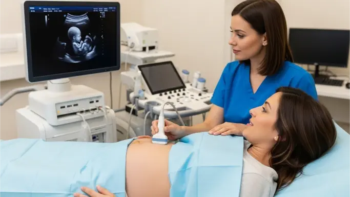
What to Expect During the Examination
The examination itself resembles standard prenatal ultrasounds. You’ll lie on an examination table while the sonographer applies gel to your abdomen and uses a transducer to obtain images and measurements. The process typically takes 20 to 30 minutes, depending on fetal position and whether additional assessments are needed.
Arriving with a comfortably full bladder can improve visualization in early pregnancy, though this becomes less important in later trimesters. Wearing comfortable clothing that provides easy abdominal access makes positioning easier.
Discussing Results
Many facilities provide preliminary information during the scan, though official results typically come through your primary obstetric provider who reviews findings in the context of your complete medical history. Don’t hesitate to ask questions during the scan or at follow-up appointments to ensure you understand what measurements mean for your specific situation.
Making Informed Decisions About Your Ultrasound Scan in Bangalore
Understanding growth scans empowers you to participate actively in pregnancy care decisions. These specialized examinations provide crucial information about fetal development, helping identify concerns early when interventions can optimize outcomes. When your healthcare provider recommends growth monitoring, it reflects appropriate attention to factors that could affect your pregnancy rather than cause for immediate alarm.
The combination of sophisticated measurement techniques, careful interpretation of growth patterns, and integration with other clinical information allows modern obstetrics to identify and manage growth abnormalities more effectively than ever before. Whether your growth scans show reassuring normal development or identify concerns requiring additional management, these examinations play a vital role in ensuring the best possible outcomes for you and your baby.
Conclusion: The Value of Growth Monitoring Through Ultrasound Scan in Bangalore
Growth scans represent an essential component of modern prenatal care for pregnancies requiring enhanced monitoring. These specialized examinations measure specific fetal body parts, calculate estimated weight, and assess the intrauterine environment to ensure babies are developing appropriately. While not every pregnancy requires serial growth scanning, women with risk factors benefit tremendously from this proactive monitoring approach. The detailed information obtained guides clinical decisions about timing and mode of delivery, helps identify complications early, and ultimately contributes to healthier outcomes. When seeking an ultrasound scan in Bangalore for growth monitoring, you’re accessing sophisticated technology and expertise that allows your healthcare team to provide personalized care tailored to your pregnancy’s unique characteristics. Understanding what growth scans measure, when they’re recommended, and how results influence care empowers you to approach these examinations with confidence rather than anxiety, knowing they represent thorough, thoughtful prenatal care designed to support the healthiest possible pregnancy journey.