enquiry@ecotowndiagnostics.com
Send Your Mail 24/7 Support
Mon - Sat 6:30AM - 10:00PM
Sunday 6:30AM - 3:00PM
Experiencing sudden, severe flank pain accompanied by nausea and blood in your urine can be alarming. These symptoms often point toward kidney stones, a common yet painful condition affecting millions worldwide. When you visit a healthcare facility for diagnosis, medical professionals typically recommend computed tomography as the gold standard for detecting these mineral deposits. Understanding what happens during a CT scan in Bangalore and why this imaging technique has become the preferred diagnostic tool can help ease anxiety and prepare you for the examination process.
Kidney stones, medically known as nephrolithiasis, form when minerals and salts crystallize in your kidneys or urinary tract. While several diagnostic methods exist, CT imaging has emerged as the most reliable and efficient option for identifying these painful obstructions. This comprehensive guide explores the accuracy, speed, and complete procedure of CT imaging for kidney stone detection.
Kidney stones vary significantly in size, composition, and location within the urinary system. Some stones measure just a few millimeters and pass naturally without intervention, while others grow large enough to block urine flow completely, requiring immediate medical attention. The chemical composition ranges from calcium oxalate and calcium phosphate to uric acid and struvite stones, each with different characteristics and treatment approaches.
Accurate diagnosis becomes crucial because treatment strategies differ dramatically based on stone size, location, and type. Small stones may only require pain management and increased fluid intake, while larger obstructions might necessitate surgical intervention or lithotripsy procedures. Without precise imaging, physicians cannot determine the optimal treatment pathway, potentially leading to unnecessary procedures or delayed care.
Traditional diagnostic methods like ultrasound and plain X-rays have limitations in stone detection. Ultrasound provides excellent real-time imaging but may miss smaller stones or those located in certain positions. Plain radiography can visualize calcium-containing stones but fails to detect radiolucent stones composed of uric acid or certain other minerals. These limitations explain why CT technology has become the preferred first-line diagnostic tool.
The superiority of CT imaging for kidney stone diagnosis stems from multiple technological advantages. Unlike other imaging modalities, computed tomography creates detailed cross-sectional images of your body, allowing radiologists to visualize stones regardless of their chemical composition. This comprehensive visualization capability makes it the most sensitive diagnostic tool available.
Studies consistently demonstrate that CT imaging achieves detection rates exceeding 95% for kidney stones of all types and sizes. This remarkable accuracy comes from the technology’s ability to differentiate between various tissue densities. Stones appear as high-density objects against the surrounding soft tissues, making them easily identifiable even when measuring just a few millimeters.
The three-dimensional reconstruction capabilities of modern CT scanners provide physicians with comprehensive spatial information. They can precisely determine stone location within the kidney or ureter, measure exact dimensions, and assess the degree of urinary obstruction. This detailed information directly influences treatment decisions and helps predict whether stones will pass spontaneously or require intervention.
When someone experiences acute renal colic, time becomes critical for both comfort and preventing complications. CT imaging for kidney stones typically requires just 5 to 10 minutes from start to finish, making it one of the fastest diagnostic procedures available. This rapid acquisition time means patients suffering from severe pain receive quick answers and can begin appropriate treatment without lengthy delays.
The efficiency extends beyond scan time. Radiologists can interpret CT images quickly, often providing preliminary results within an hour. This rapid turnaround enables emergency departments to make swift treatment decisions, whether that involves pain management, scheduling procedures, or determining safe discharge home with follow-up care instructions.
Proper preparation ensures optimal image quality and a smooth examination experience. Fortunately, CT imaging for kidney stones requires minimal preparation compared to other diagnostic procedures, making it particularly convenient for emergency situations.
Most facilities performing kidney stone CT scan in Bangalore do not require fasting or special dietary restrictions. You can typically eat and drink normally before your appointment, which differs from abdominal CT scans that might require contrast material. However, you should inform the medical staff about any medications you’re taking, especially blood thinners or diabetes medications.
Remove all metal objects including jewelry, watches, belts with metal buckles, and eyeglasses before the scan. Metal creates artifacts on images that can obscure important details. Most facilities provide lockers for secure storage of personal belongings during the examination.
If you’re pregnant or suspect you might be pregnant, inform the technologist immediately. While CT imaging provides crucial diagnostic information, physicians weigh the benefits against potential radiation risks during pregnancy and may consider alternative imaging methods when appropriate.
Standard kidney stone CT scans typically use a non-contrast protocol, meaning no intravenous dye injection is necessary. This approach reduces procedure time, eliminates concerns about contrast allergies, and provides excellent stone visualization. The absence of contrast material also makes the examination suitable for patients with kidney dysfunction who cannot safely receive contrast agents.
However, some clinical situations may warrant contrast-enhanced scanning to evaluate complications like infection, obstruction severity, or anatomical variations. Your physician determines whether contrast administration is necessary based on your specific symptoms and medical history.
Understanding what happens during the examination helps reduce anxiety and ensures you can cooperate effectively with positioning requirements.
Upon arriving at the imaging center, you’ll complete registration paperwork and provide relevant medical history. The staff reviews your symptoms, previous imaging studies, and any contraindications to scanning. This screening process typically takes 10 to 15 minutes before you proceed to the examination area.
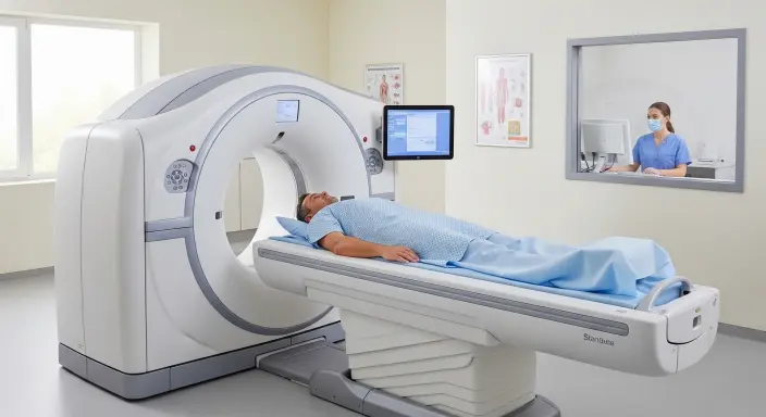
The CT technologist positions you on a motorized table, typically lying flat on your back with arms raised above your head. This position provides unobstructed visualization of your abdomen and pelvis. Pillows or positioning devices may be used to ensure comfort and proper alignment. The technologist explains breathing instructions, as remaining still and following breath-hold commands ensures optimal image quality.
The table slowly moves through the circular CT scanner opening, which resembles a large donut. Modern scanners are wider and shorter than older models, reducing claustrophobic feelings. The actual scanning takes just 10 to 30 seconds during which you must remain completely still. You’ll hear mechanical whirring sounds as the scanner’s components rotate around you, but the process is painless and non-invasive.
Throughout the scan, the technologist monitors you from an adjacent control room through a window and intercom system. They can see and hear you constantly, and you can communicate if you experience any discomfort or concerns. After the brief scan completes, the table returns to its starting position and you can immediately resume normal activities.
One concern patients often raise involves radiation exposure from CT imaging. While computed tomography does use X-rays, and therefore involves radiation exposure, the diagnostic benefits typically far outweigh the minimal risks, especially in acute situations requiring immediate diagnosis.
Modern CT scanners incorporate dose reduction technologies that minimize radiation while maintaining diagnostic image quality. Low-dose protocols specifically designed for kidney stone detection deliver significantly less radiation than standard abdominal CT examinations. These protocols recognize that stone detection requires less radiation than soft tissue evaluation.
The radiation dose from a kidney stone CT scan is comparable to the natural background radiation exposure accumulated over several months to a few years, depending on where you live. For patients with recurrent kidney stones requiring multiple scans over their lifetime, physicians carefully weigh the necessity of each examination and may alternate with ultrasound when clinically appropriate.
After completing the scan, a radiologist—a physician specializing in medical imaging interpretation—analyzes the images and prepares a detailed report. This report describes any stones detected, including their size, location, density measurements, and associated complications like hydronephrosis or ureteral obstruction.
The radiologist’s report provides crucial information that guides treatment decisions. Stone size, measured in millimeters, helps predict the likelihood of spontaneous passage. Stones smaller than 5mm typically pass naturally with conservative management, while larger stones often require intervention. Location matters equally—stones in the kidney may cause different symptoms than those lodged in the ureter.
Density measurements, expressed in Hounsfield units, can sometimes suggest stone composition. Calcium-containing stones appear brighter and denser than uric acid stones. This information helps physicians predict which medications might dissolve certain stone types or prevent future formation.
Your physician reviews the CT results and discusses appropriate treatment options. Small stones with minimal obstruction may only require pain management, increased fluid intake, and medications to facilitate passage. Larger stones or those causing significant obstruction might need procedures like extracorporeal shock wave lithotripsy, ureteroscopy, or percutaneous nephrolithotomy.
The detailed anatomical information from CT imaging helps surgeons plan minimally invasive procedures with greater precision, potentially reducing operative time and improving outcomes. Follow-up imaging, often using ultrasound to minimize radiation exposure, monitors treatment effectiveness and confirms complete stone clearance.
While CT imaging for kidney stones is safe for most individuals, certain populations require special considerations or alternative approaches.
Children experiencing suspected kidney stones present unique challenges. While CT scan in Bangalore provides excellent diagnostic accuracy, physicians carefully consider radiation exposure in growing bodies. Many pediatric centers use ultrasound as the initial imaging modality, reserving CT for cases where ultrasound proves inconclusive or when precise stone characterization is essential for treatment planning.
When CT becomes necessary for pediatric patients, technologists use specialized low-dose protocols designed specifically for children’s smaller body sizes. These protocols maintain diagnostic quality while minimizing radiation exposure to the lowest level possible.
Pregnancy creates special diagnostic challenges because standard imaging modalities carry theoretical risks to the developing fetus. Ultrasound becomes the preferred first-line imaging tool for pregnant women with suspected kidney stones. However, when ultrasound proves inadequate and the clinical situation demands definitive diagnosis, CT may be performed using modified protocols that minimize radiation exposure to the pelvis.
Physicians carefully document the medical necessity when ordering CT examinations during pregnancy, ensuring the diagnostic benefits justify any theoretical risks. Modern shielding techniques and dose reduction protocols further minimize fetal exposure.
Understanding how CT imaging compares to other diagnostic options helps patients appreciate why physicians prefer this modality for kidney stone evaluation.
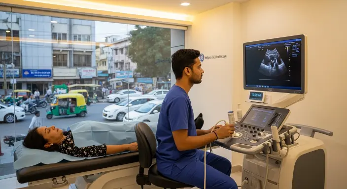
Ultrasound provides radiation-free imaging and works well for detecting kidney stones and hydronephrosis, but it struggles to visualize stones in the mid-ureter and may miss smaller stones. The examination quality depends heavily on operator skill and patient body habitus, introducing variability in diagnostic accuracy.
Plain abdominal radiography, historically used for kidney stone screening, misses radiolucent stones entirely and provides limited anatomical detail. While inexpensive and widely available, its poor sensitivity makes it inadequate for comprehensive evaluation.
Intravenous pyelography, once considered the gold standard, requires contrast injection, takes significantly longer to complete, and provides less detailed information than modern CT imaging. Most facilities have replaced this outdated technique with computed tomography for kidney stone diagnosis.
Modern CT technology continues evolving, improving both diagnostic capabilities and patient comfort. Iterative reconstruction algorithms reduce image noise, allowing lower radiation doses while maintaining or improving image quality. Wider scanner openings accommodate larger patients more comfortably and reduce claustrophobic reactions.
Artificial intelligence applications are emerging to automatically detect and measure kidney stones, potentially speeding diagnosis and reducing radiologist workload. These advanced software tools may soon provide instant preliminary results, further reducing the time from symptom onset to treatment initiation.
When facing the possibility of kidney stones, understanding your diagnostic options empowers you to participate actively in healthcare decisions. CT imaging provides the most accurate, fastest, and most comprehensive evaluation available for this painful condition. The brief, painless procedure delivers definitive answers that guide appropriate treatment, whether that means conservative management or surgical intervention.
The minimal radiation exposure from modern low-dose protocols presents negligible risk compared to the diagnostic benefits, especially when experiencing acute symptoms requiring immediate evaluation. Most patients complete the entire process—from arrival to departure—within an hour, making it remarkably convenient even during emergency situations.
CT imaging has revolutionized kidney stone diagnosis, offering unparalleled accuracy and speed that directly benefit patients suffering from this common condition. The technology’s ability to detect stones regardless of composition, precisely measure dimensions, and identify complications makes it the undisputed gold standard for evaluation. When you seek a CT scan in Bangalore for suspected kidney stones, you’re accessing sophisticated technology that provides physicians with the detailed information necessary for optimal treatment decisions. The combination of rapid results, comprehensive visualization, and minimal preparation requirements explains why this imaging modality has become the preferred first-line diagnostic tool worldwide. Understanding what to expect during the examination process helps reduce anxiety and ensures you can cooperate effectively, contributing to the highest quality diagnostic images possible.